Mallow, iris, orchid
Scientific plant images over the centuries

The aesthetics of scientific representations of plants and flowers are very appealing. Now, as ever, domestic and exotic plants seem within our reach thanks in part to perfect illusionistic representations. As a result of technical advances, drawings, photography, microscopy and computer-aided visualisations enable the beauty of botanic details to be seen in ever more facets.
In the history of ETH Zurich and the documents and images of ETH Library, you can find countless traces revealing the development of scientific representation over time. They show the great influence originators, customers and users have until now had on our general understanding of nature. The same factors are also mirrored in present-day professional education of scientific illustrators. One example is the Master of Knowledge Visualization degree offered by the Zurich University of the Arts (ZHdK).
Since the Renaissance, botany has continuously expanded its repertoire of visualisation techniques, which are now used side by side. The precursors of modern technical procedures like photography, microscopy and 3D visualisations were drawings executed by hand.
Herbal books of the Renaissance – In search of the ideal representation
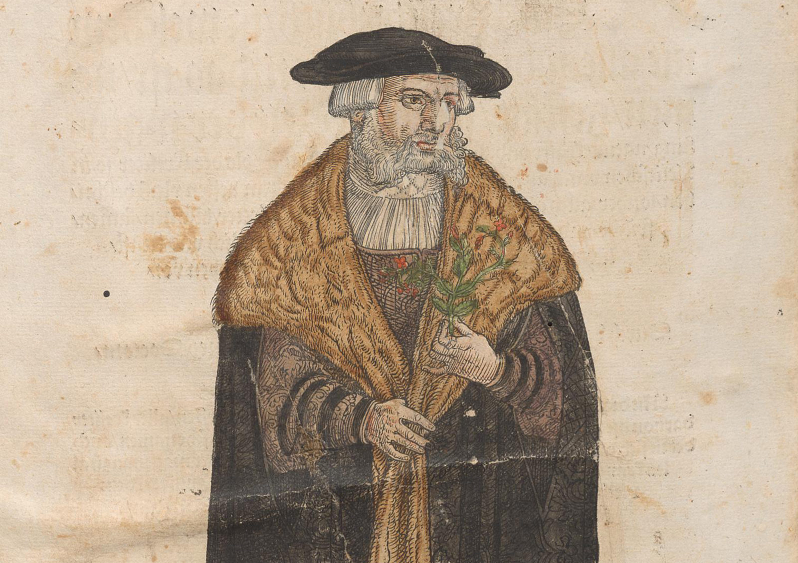
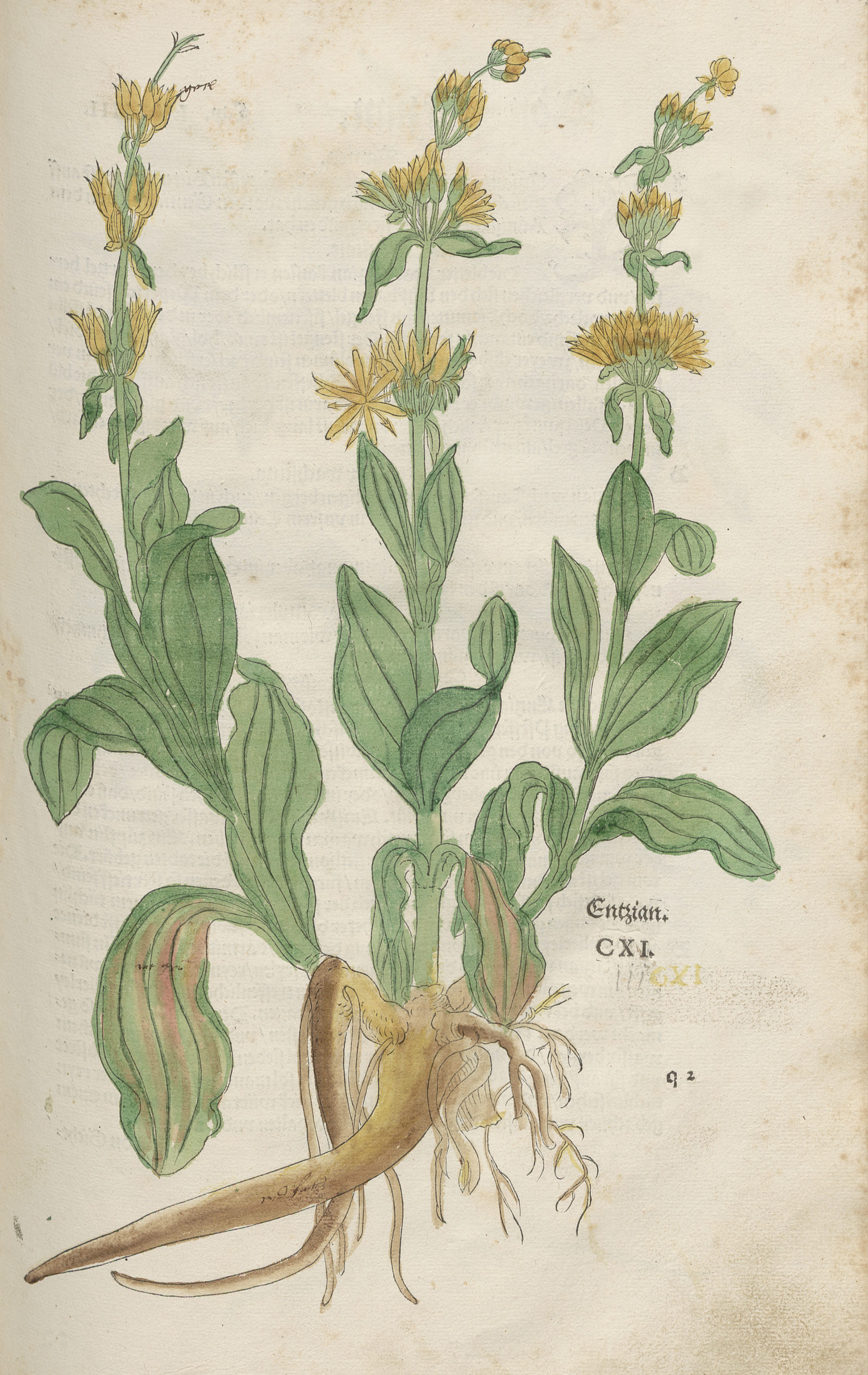
The invention of book printing provided Renaissance physicians like Leonhart Fuchs and Otto Brunfels with new opportunities. On the basis of ancient texts, they successfully published sixteenth-century knowledge of medicinal plants in what were termed herbal books and in doing so promoted botany.
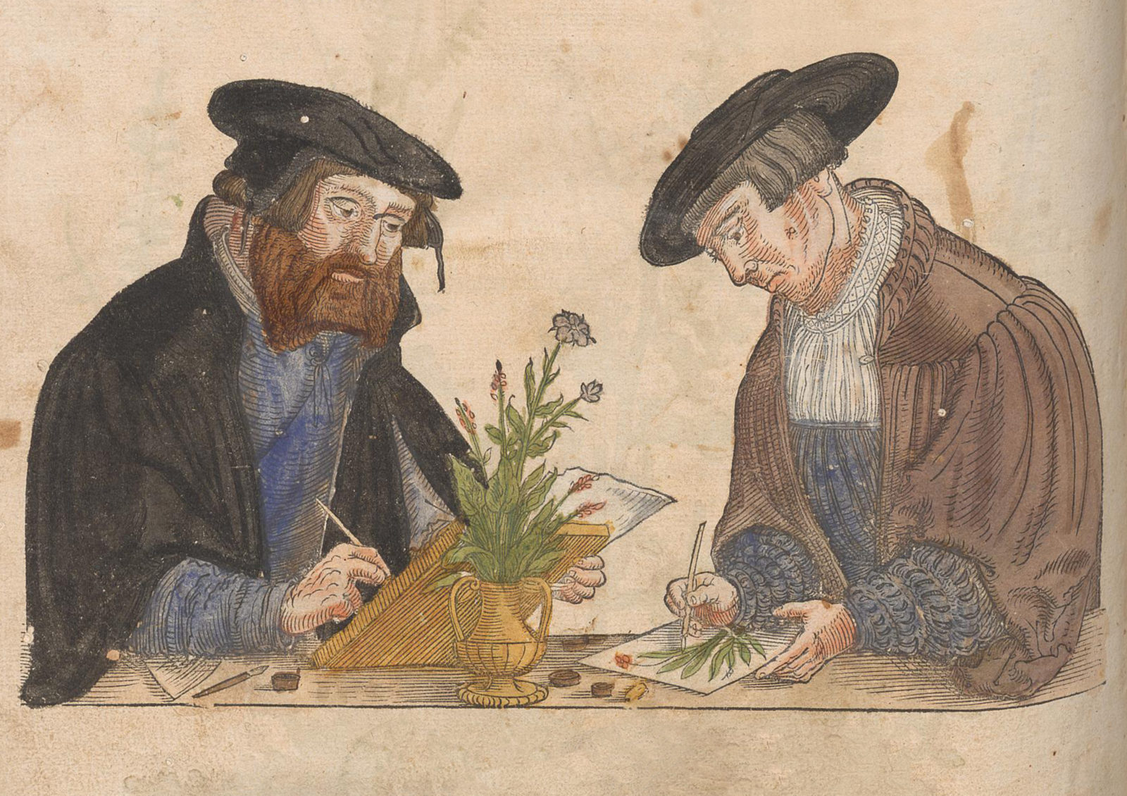
In these books the ‘fathers of botany’ systematically supplemented the textual descriptions of plants with illustrations. But this practice was not without its detractors: one of them was the German physician Janus Cornarius. He pointed out that a depiction merely showed a specific plant at a specific time in a specific place. This was not, he said, appropriate for a universal description of an entire species.
Since then, plant species have been represented in scientific illustrations in an ideal format achieved by abstraction and construction. What is shown is not simply an individual plant but an abstracted – though lifelike – “absolute” picture.
As a result of the technique of woodcuts, however, this envisaged naturalness of plant representation in the herbal books of the Renaissance was subject to strict limits.
Leonhart Fuchs’ first book of herbs, published in 1542 under the title De Historia Stirpium commmentarii insignes, featured over 500 plant illustrations. Written in Latin, it was aimed at an educated specialist readership. The popular German version of the New Kreuterbuch was published in 1563. A few copies, including ETH-Bibliothek’s, were hand-coloured.
Garden books – Impressive volumes for wealthy sponsors
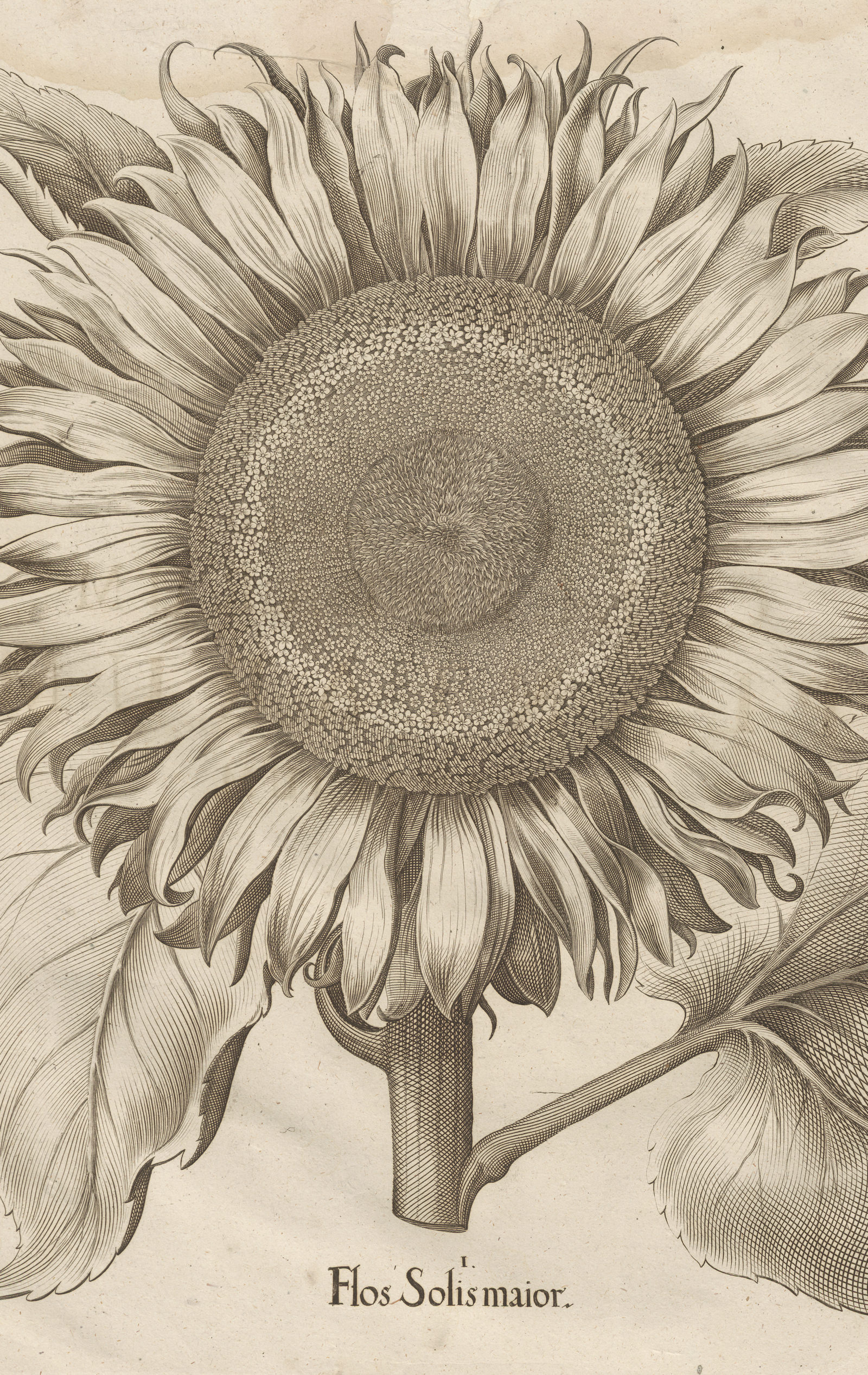
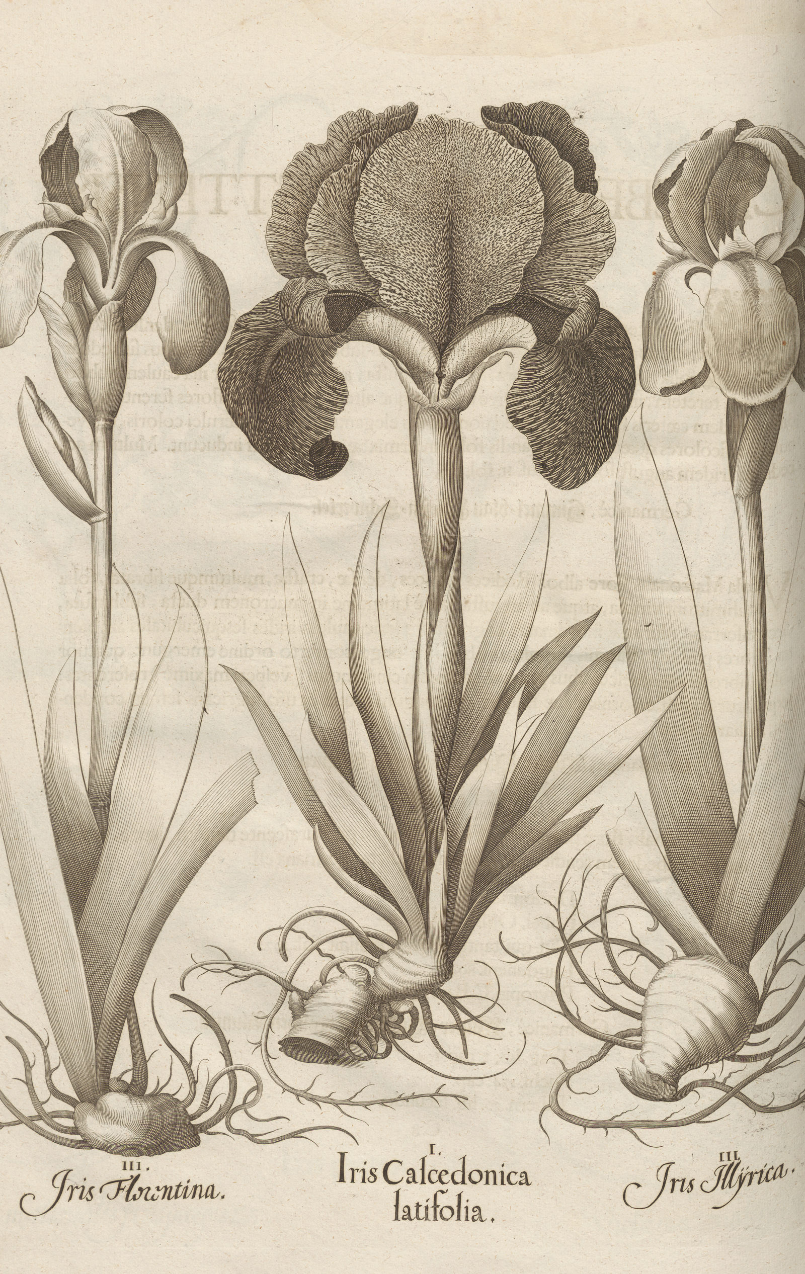
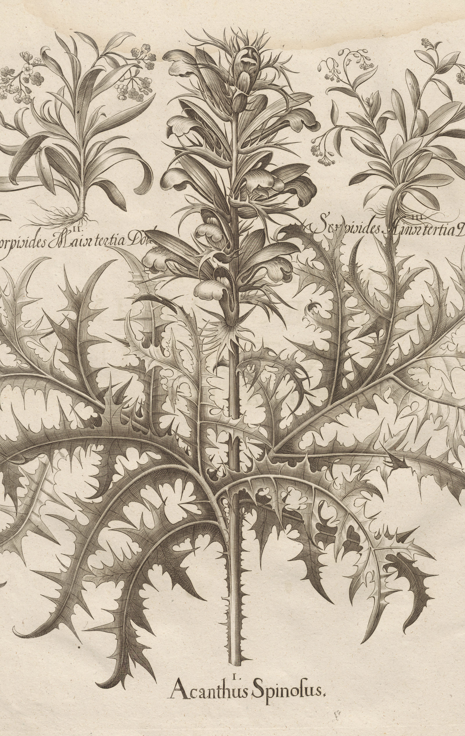
Thanks to engravings, much more detailed representations could soon be made and printed. But the ever greater expenditure for illustrators, engravers and printers also led to higher production costs for illustrated botanical works. Countless impressive volumes were created and published only with the support of wealthy sponsors.
These included ‘Hortus Eystettensis’ by Basilius Besler, which came out in 1613. This book shows over 1,000 European and exotic cultivated plants from the representative garden that prince-bishop Johann Konrad laid out around 1600 at the castle of Willibaldsburg near Eichstätt in Germany. The prince-bishop also paid for the publication, which did much to raise awareness of his garden and ran to several editions.
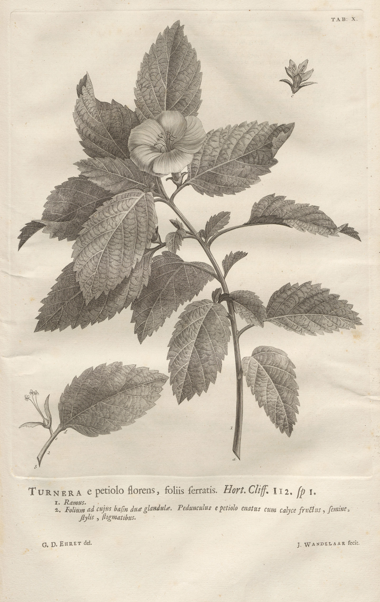
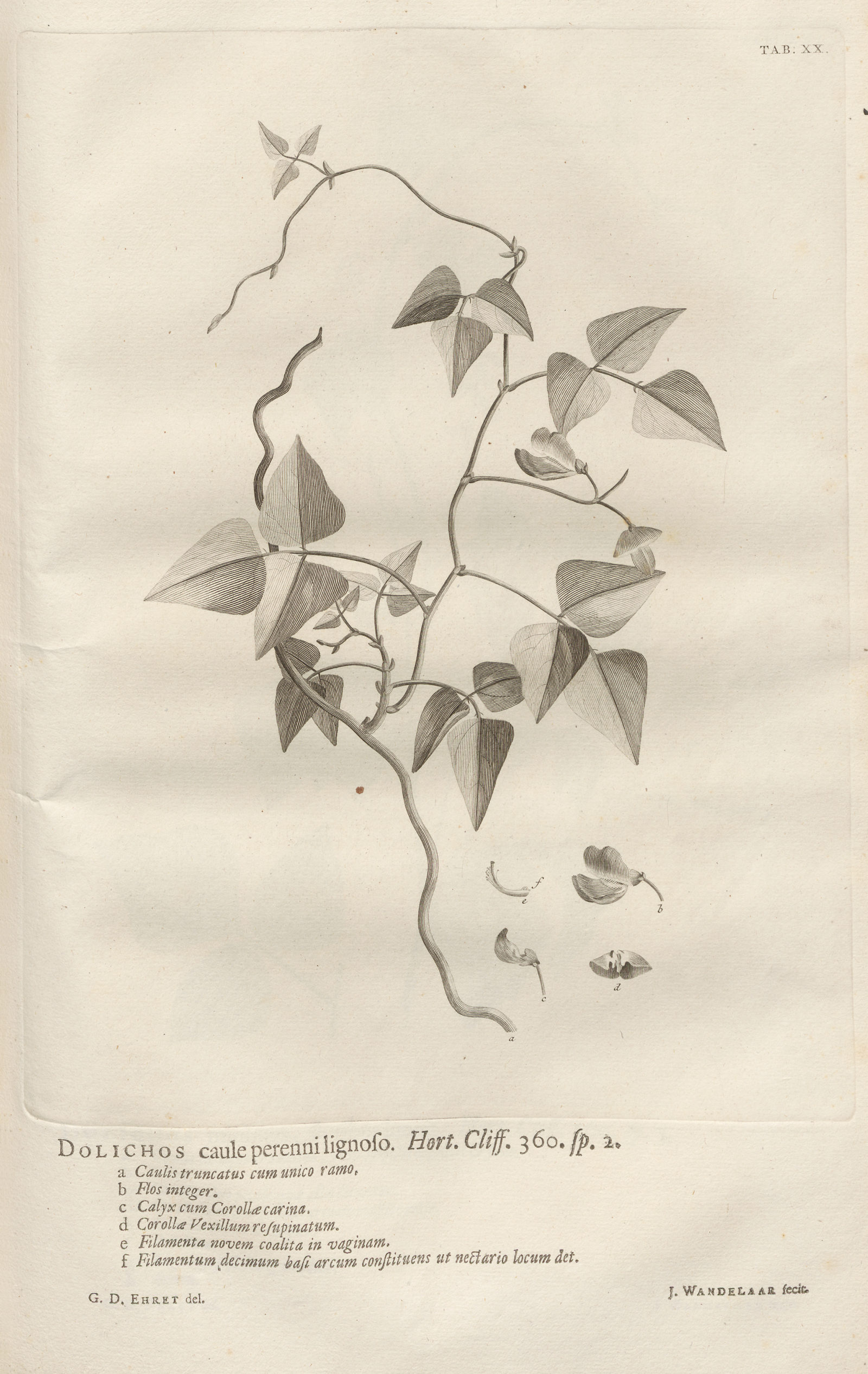
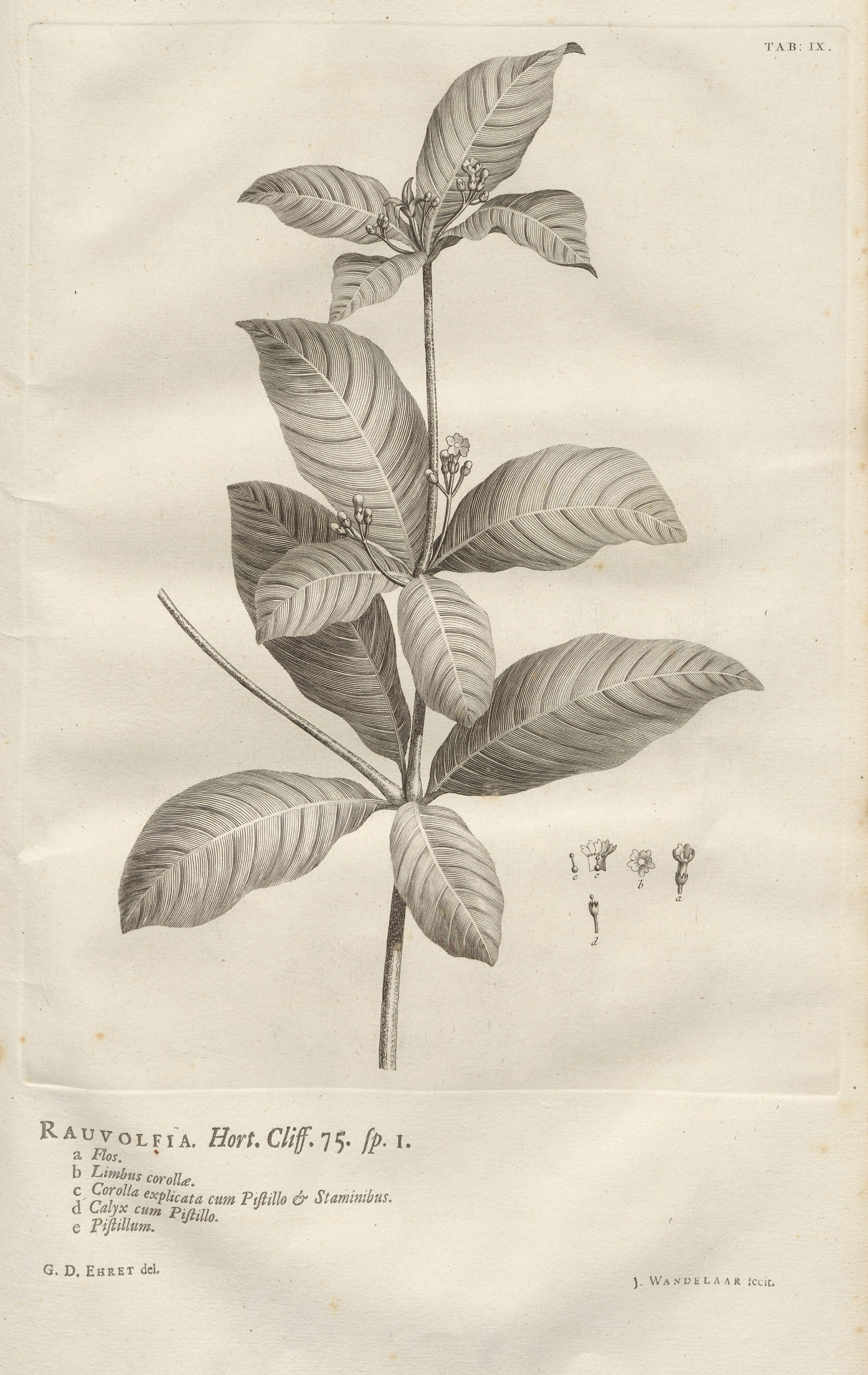
Another garden, the ‘Hortus Cliffortianus’ in Hartekamp in the Netherlands, gave its name to a work by Carl Linnaeus that came out in 1737. In it, the famous natural scientist catalogued the domestic and exotic plants in the garden and herbarium of his sponsor George Clifford. The clear separation made by Linnaeus between naming and classification of plants is still used in biology to this day.
Carl von Linnaeus’s detailed classification placed high demands on the precision of scientific illustrations. He therefore cooperated with, among others, Georg Dionysius Ehret, one of the most successful plant painters of the eighteenth century.
The flora of entire countries in classification systems
Influenced by Linnaeus’s work, classification systems were used not only to describe collections of plants artificially collected in gardens but also to systematically classify and illustrate the flora of entire countries.
Thus Niklaus Joseph von Jacquin, director of the Vienna University botanical garden and the imperial gardens around Schönbrunn Palace, published the five-volume work ‘Florae austriacae’ between 1773 and 1778. 1 Five hundred costly colour plates illustrate this flora of the Duchy of Austria.
From 1837 the botanist and zoologist Ludwig Reichenbach published ‘Deutschlands Flora: mit höchst naturgetreuen, charakteristischen Abbildungen aller ihrer Pflanzen-Arten in natürlicher Grösse und mit Analysen auf Kupfertafeln’, also running to several volumes but based on its own classification. 2
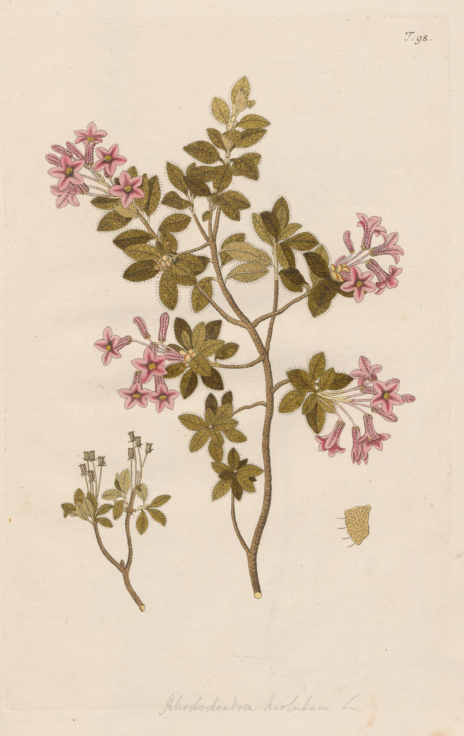
Compared with these and other extremely expensive national epic works from the heyday of illustrated botanical works, the small-format ‘Sammlung von Schweizer Pflanzen’, which came out in 1840, looks rather modest. 3 The lithographer Jonas David Labram published the first partial deliveries of this work by himself. But he was soon assisted by the botanist and doctor Johannes Hegetschweiler.
The beauty of Swiss apples and pears
Even though the most expensive illustrated works appeared in other countries, botanical works were also commissioned in Switzerland that had a national orientation, scientific claim and high-quality illustrations. An example is the two-volume ‘Schweizerische Obstsorten’, published by the Swiss Agricultural Association between 1863 and 1872.
The preface to this publication of Swiss varieties of apples and pears and the characteristics of their visual and textual description also sets out the requirement criteria for illustrations and text:
The most natural illustrations possible […] represent in their actual size:
- The side view of a mesocarp with calyx, seen from the side facing the sun.
- A longitudinal section […].
- A mature, leafed branch […]. The course of the leaf vein network […] must be shown precisely on a branch leaf in accordance with a photographic record.
The brief text, as accurate as possible, contains:
— ‘Schweizerische Obstsorten’, 1863
- The systematic or […] the local name of the species […].
- The origin and distribution of the species in Switzerland.
- A description of the shape and size of the fruit […].
- Details on how the fruits are used. 4
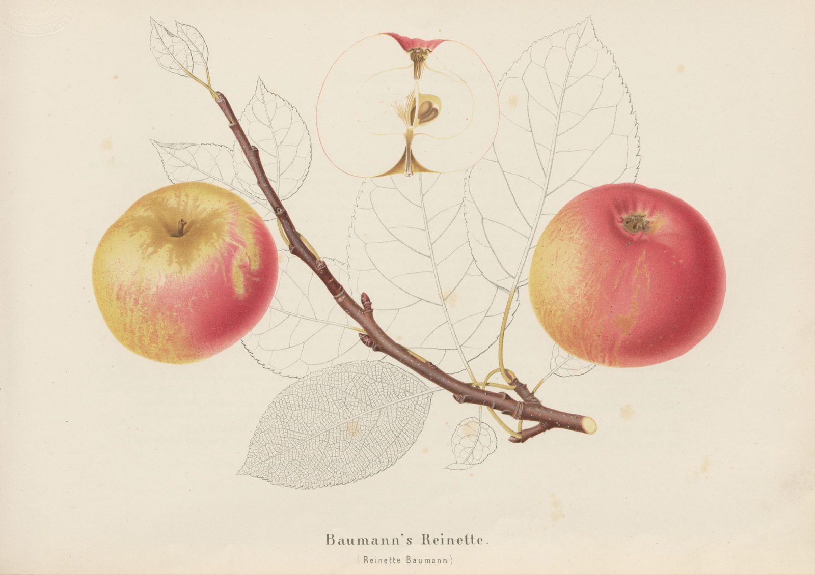
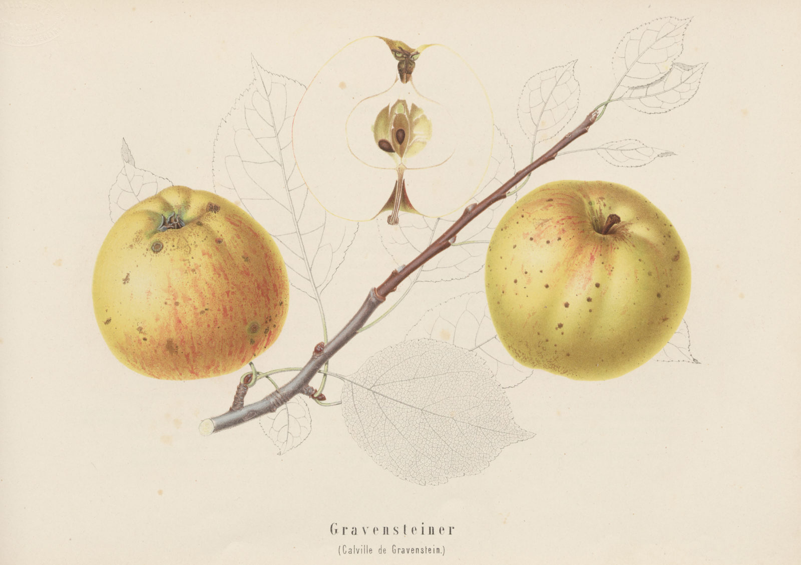
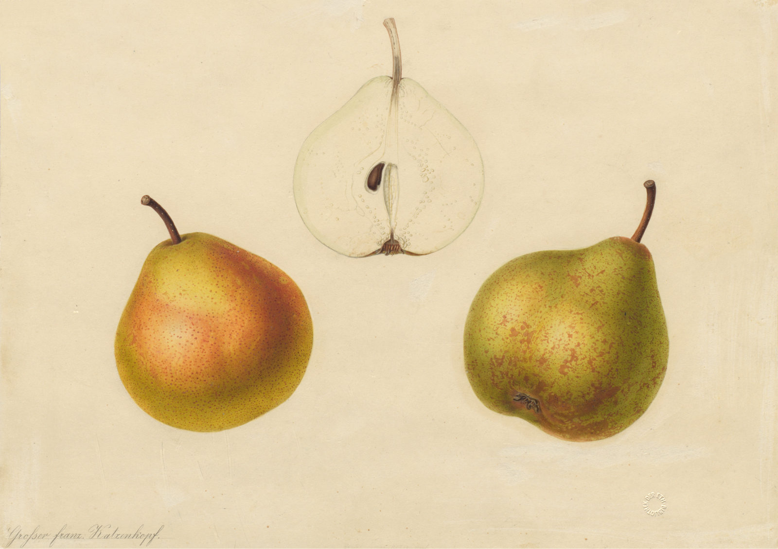
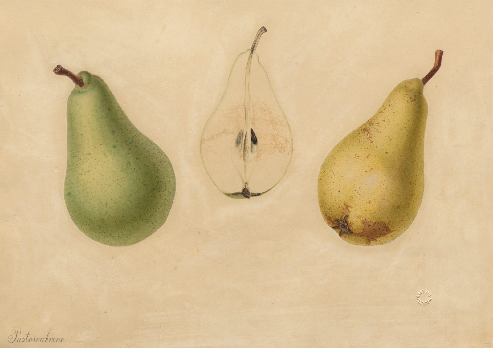
The association commissioned the Swiss painter and illustrator Salomon Bühlmeier (1814–1876) to execute the drawings. The publication was assisted by a committee set up for this purpose under the leadership of Johann Jakob Kopp, professor of forestry at the then Federal Polytechnic, now ETH Zurich.
It is probably thanks to this link that the original drawings were handed over to ETH Zurich when the project was completed. 5 This was in keeping with the widespread tradition of donating original scientific drawings to museums, botanical gardens and universities so that they could be made accessible to science and be preserved for posterity.
Switzerland’s prehistoric plant world – From fossil to oil painting
While Professor Kopp provided scientific assistance with preparing an inventory of contemporary Swiss types of fruit, two of his colleagues – the botanist Oswald Heer and the geologist Arnold Escher von der Linth – turned their attention to the plants of the past, researching the prehistoric flora of Switzerland.
The fossils that had been discovered in Öhningen in southern Germany played a vital role in this respect. In the eighteenth century these fossil finds were still being interpreted as witnesses of the Flood. In the middle of the nineteenth century, however, Heer and Escher systematically looked for fossils in Öhningen, created a collection and evaluated them scientifically.
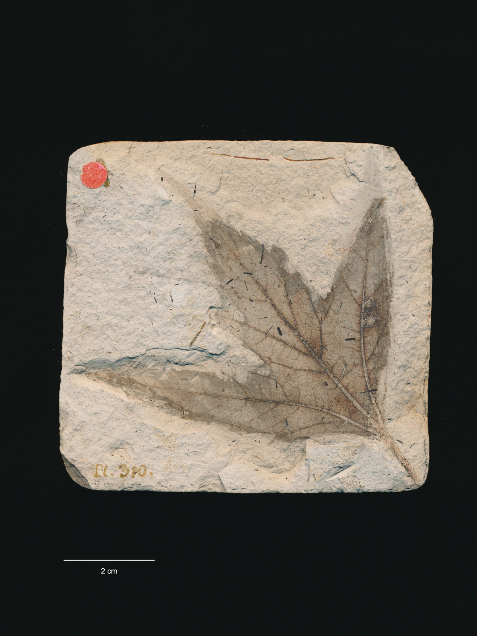
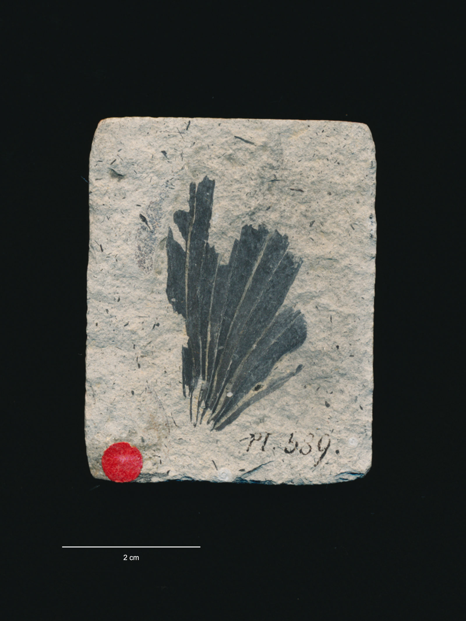
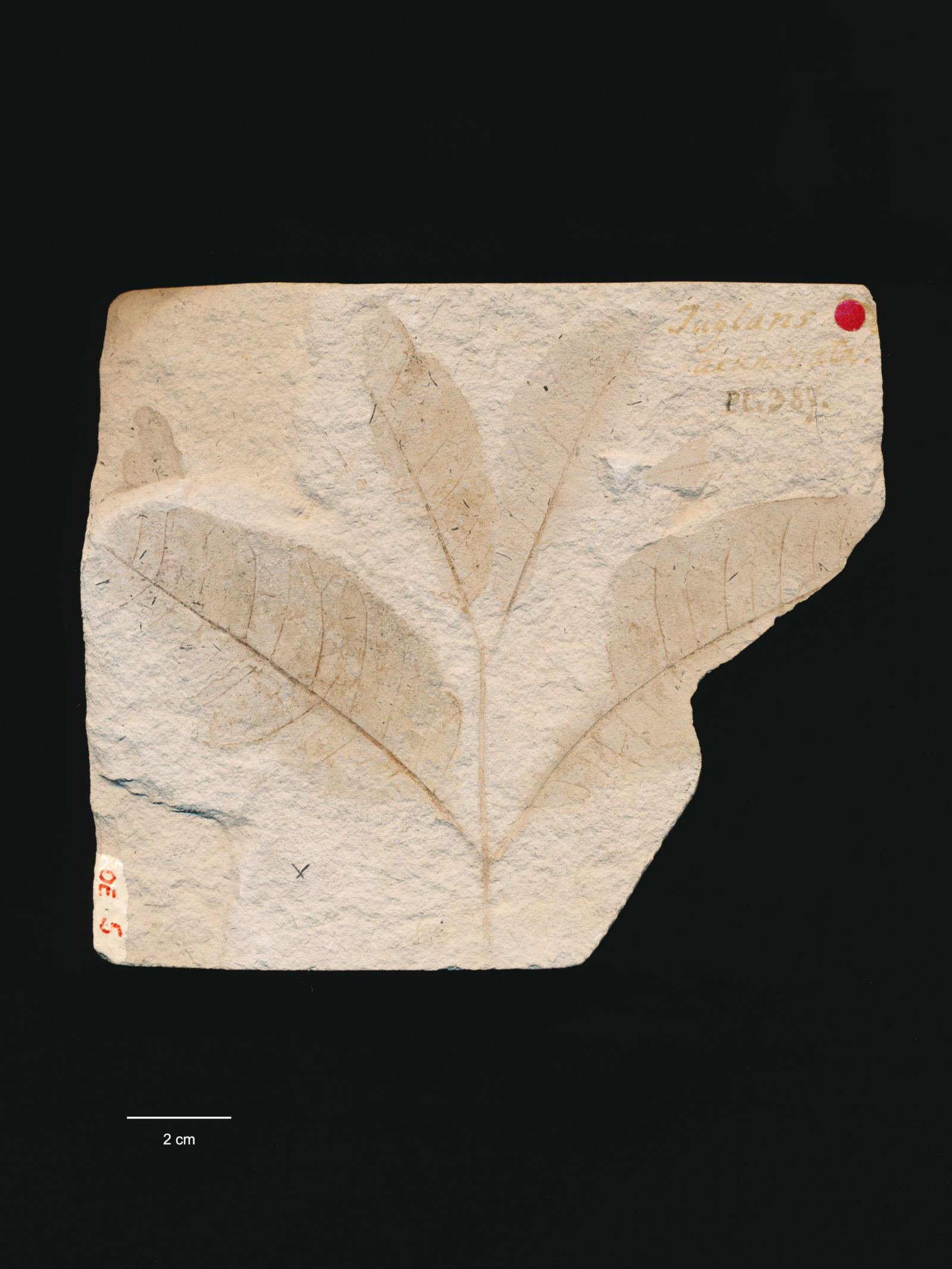
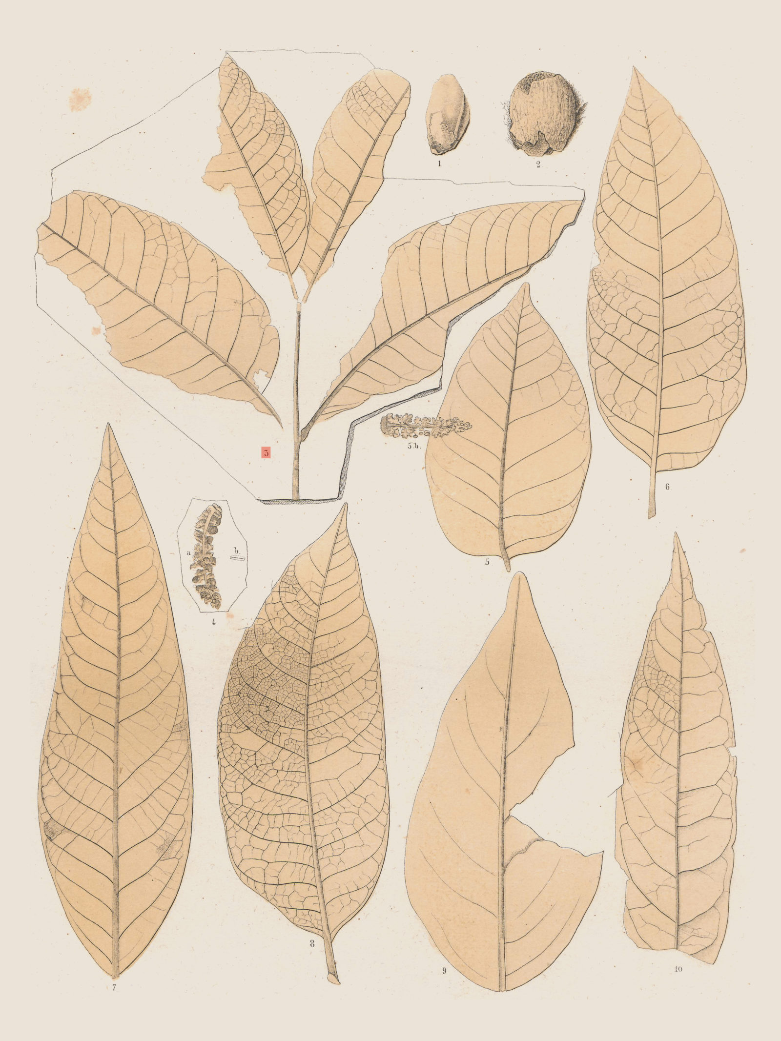
Heer published the main results in the three-volume work ‘Flora tertiaria Helvetiae. Die tertiäre Flora der Schweiz’ (1855–1859), in which numerous plant fossils were documented graphically in over 150 plates. Heer explicitly stated why he had decided to use drawings and not photography as the method of representation:
Only a handful of the images were prepared by me; most were executed by a skilled draughtsman, Mr Brugier, in my room and under my specific supervision. […] I could not use photography and nature printing for my purpose. Photography does not provide the finer veins, nor does it provide the necessary sharpness, and nature printing often lets us down where contours and veins are indicated only by delicate, […] lines […]. Furthermore, with both representation methods leaves are often hidden in diverse ways and not infrequently the image is confused and blurred by objects that are wholly foreign to the plant. With hand drawings, all these random formations can be done away with and instead what is characteristic can be highlighted more sharply […]. 6
— Oswald Heer, 1855
But visualisation of the Öhningen finds subsequently went well beyond the work of a ‘skilled draughtsman’. The scientific results from the Öhningen plant and animal world were so dense that they invited an overall visual reconstruction.
The painter Adolf Rudolf Holzhalb achieved this in his large-scale oil painting ‘Öhningen im Miozän vor etwa 13 Millionen Jahren’ of 1871.
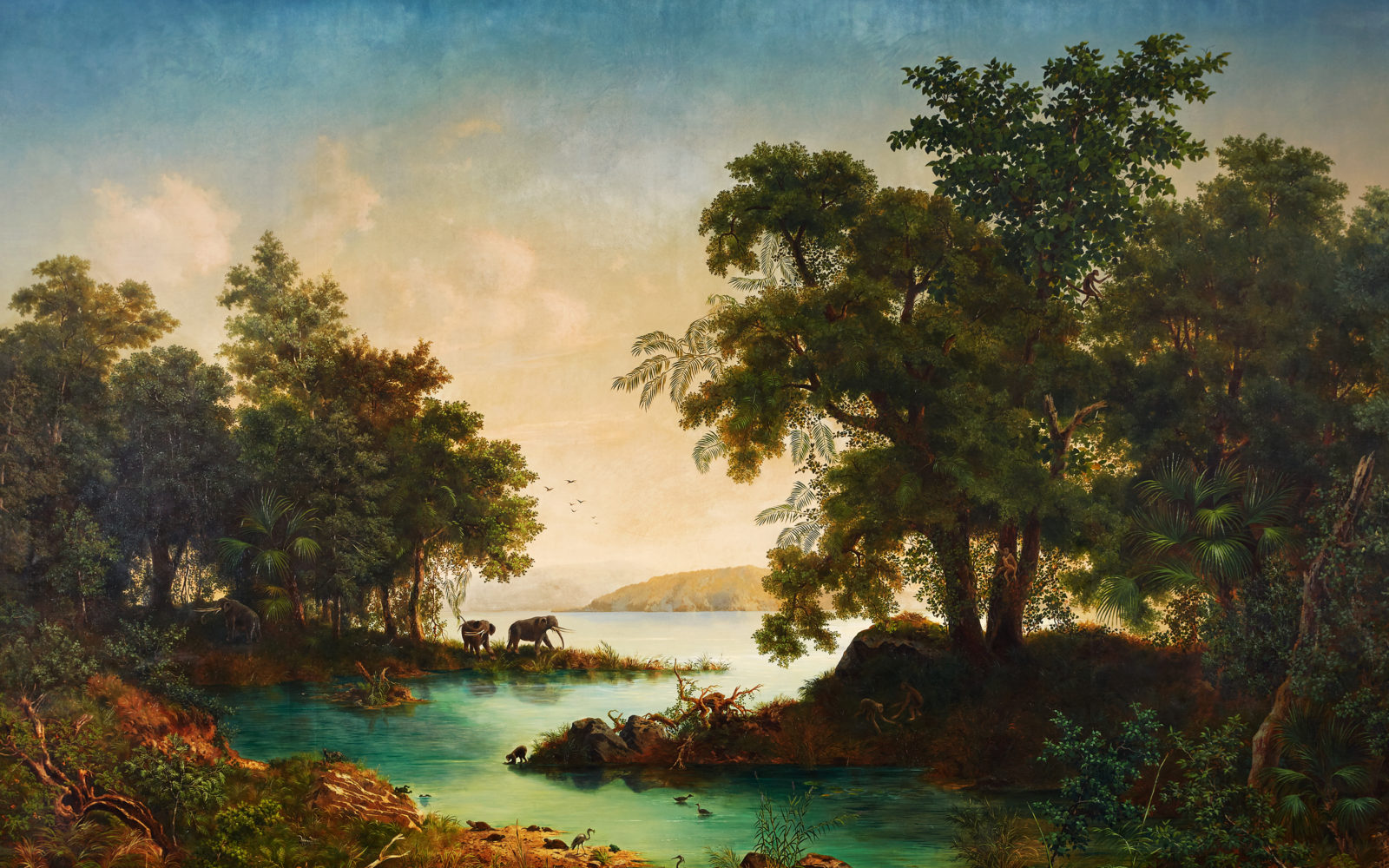
The work performed by Heer and Escher von der Linth led not to a fantastic primeval ideal landscape but instead to an educational picture underpinned by the state of science at the time. In the painting, now on display in the atrium of the NO building at ETH Zurich 7 , it is possible for example to identify fan palms and many other plants and to associate them with and examine them alongside the fossils still held today in the Geological Collections. But artistic freedom was in no way lost in the process. This is what Heer says about the striking mastodons in the centre of the picture:
But the most powerful animal of the time, the elephant-like mastodon (Mastodon tapiroides), also appears in the Öhningen groves. The artist has two of them approaching the water on a headland while a third retreats into the forest undergrowth. 8
— Oswald Heer, 1871
Photography or drawing? – A long-standing discussion
Not long after its invention in the nineteenth century, the medium of photography challenged the long tradition of graphic plant illustration. Oswald Heer’s achievements also show this. The question arose as to whether photography could replace drawings, with their demands on time.
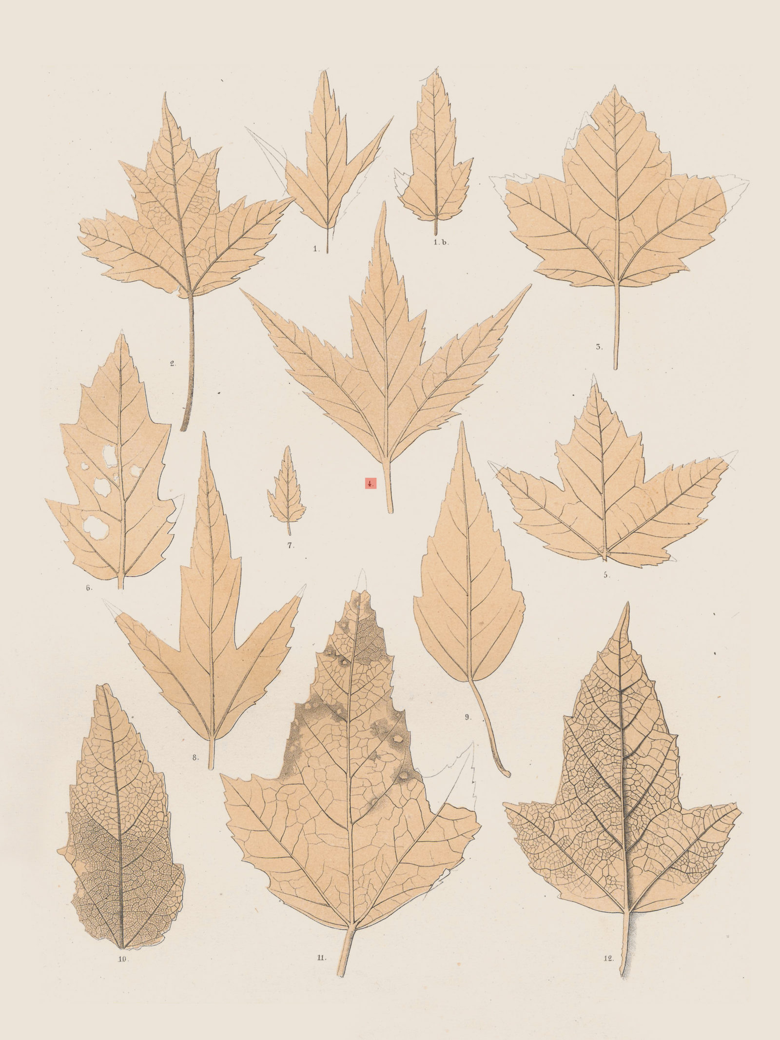
Photography is at a disadvantage when generalised features detached from the individual plant need to be expressed visually. This is what the two ETH professors, Hans Ernst Hess and Elias Landolt, said in their three-volume standard work ‘Flora der Schweiz und angrenzender Gebiete’, running to over 2,500 pages:
Ms Rosmarie Hirzel prepared the drawings from herbal material and living plants. They are an important component of our “Flora”; this is because good drawings reflect the external features of a species better and more clearly than comprehensive descriptions. Only where the characteristic features cannot be grasped by line drawings (colours, lustre) are illustrations done away with […] it is also often details of importance to classification (e.g. fruits, hairiness) that are illustrated as enlargements. […] Much of what is in the drawings is schematised in textbook fashion: Whatever is significant is highlighted. To achieve the same objective using photos would have incurred intolerable expense. 9
— Hans Ernst Hess, Elias Landolt, 1976
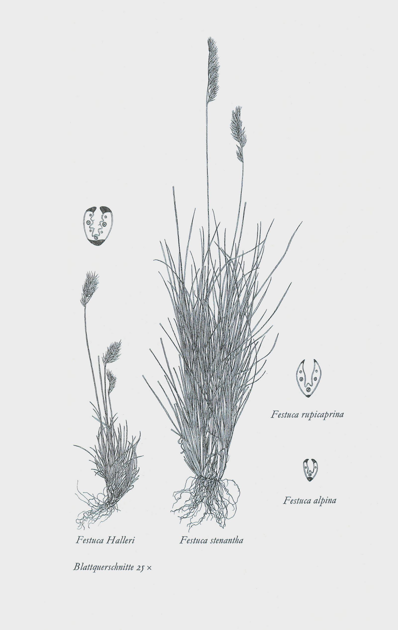
The same monochrome line drawings now stored in the ETH Zurich archives were also used for ‘Bestimmungsschlüssel zur Flora der Schweiz und angrenzender Gebiete’ (H E Hess, E Landolt, M Hirzel, M Baltisberger), which is popular among experts and suited to field studies. The seventh edition of this standard work came out in 2015. Their didactic clarity meant that the same drawings were included in the textbook on ‘Systematic botany’ also used in ETH Zurich. 10 In parallel with photography, drawings have therefore successfully held their ground as scientific illustrations to this day.
But photography has of course long since found its way into the domain of scientific plant illustration. This is shown for example by the photo inventory of the ETH Zurich Geobotanical Institute covering the period from 1880 to around 1960 in the ETH Library Image Archive: Biologists were intensively using photography early on to record images of plants from all over the world.
Photography seems to be most efficient where it is used to document a plant family as comprehensively as possible in image form. An example of this is the documentation of European orchids by Hans R Reinhard (1919–2007). From the 1960s Reinhard undertook a number of research trips through Europe as a side line during which he took more than 20,000 photos of orchids. He usually photographed several individual plants of a particular species to create a broad image base that was as representative as possible. This resulted in images of the entire orchid as well as extreme close-ups of details.
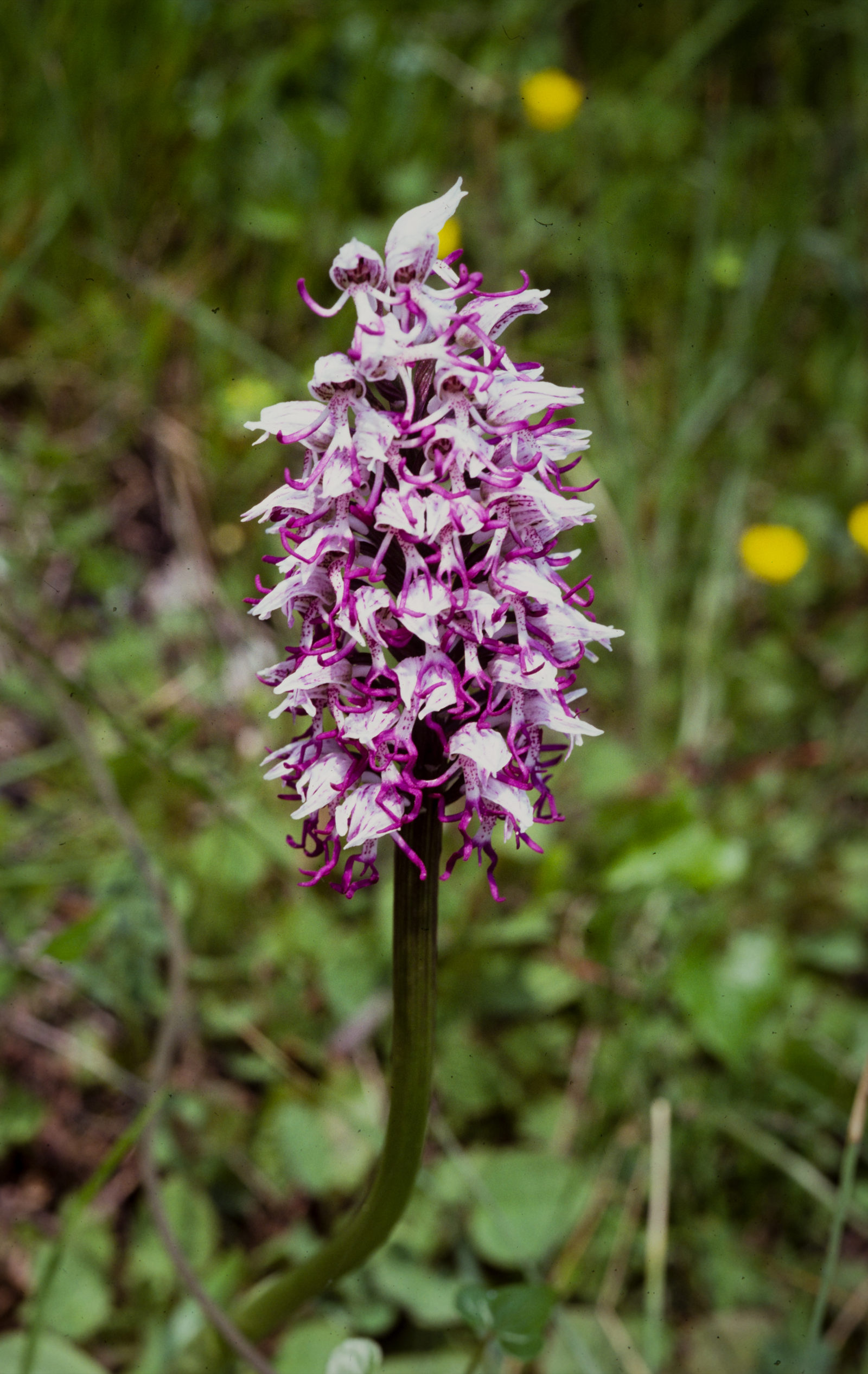
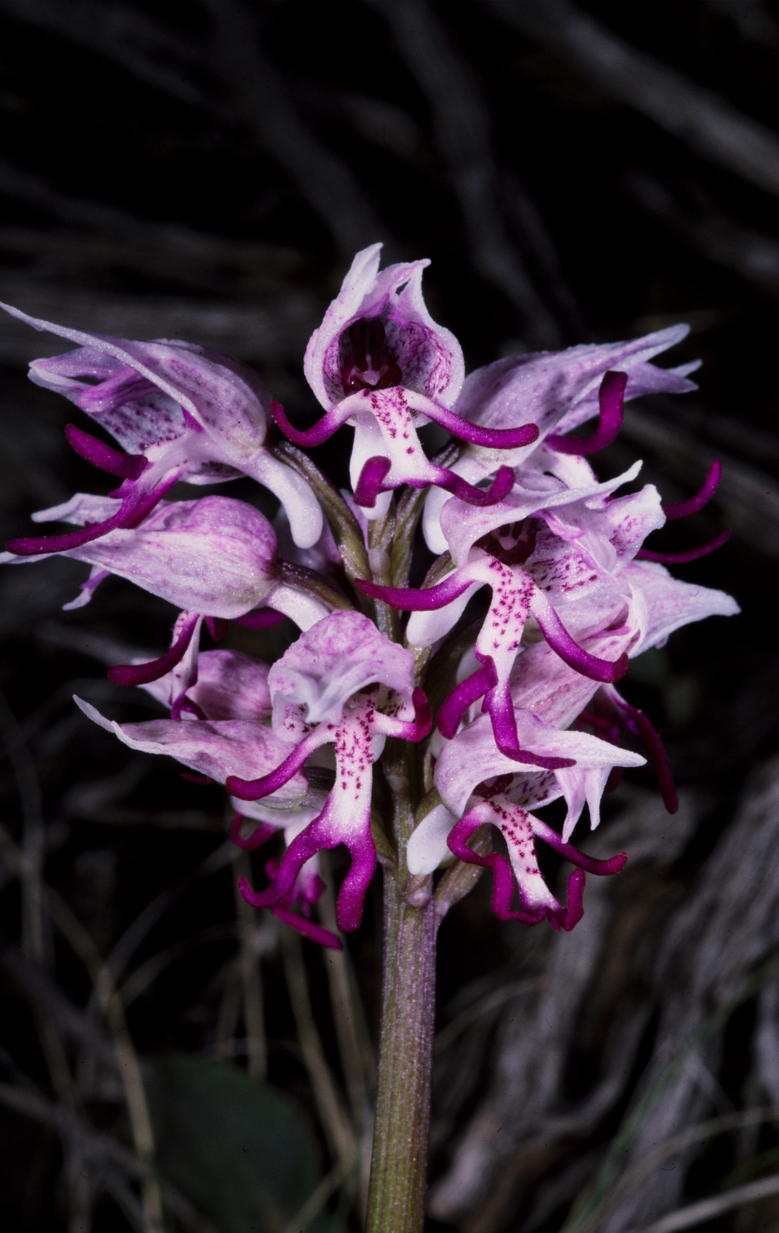
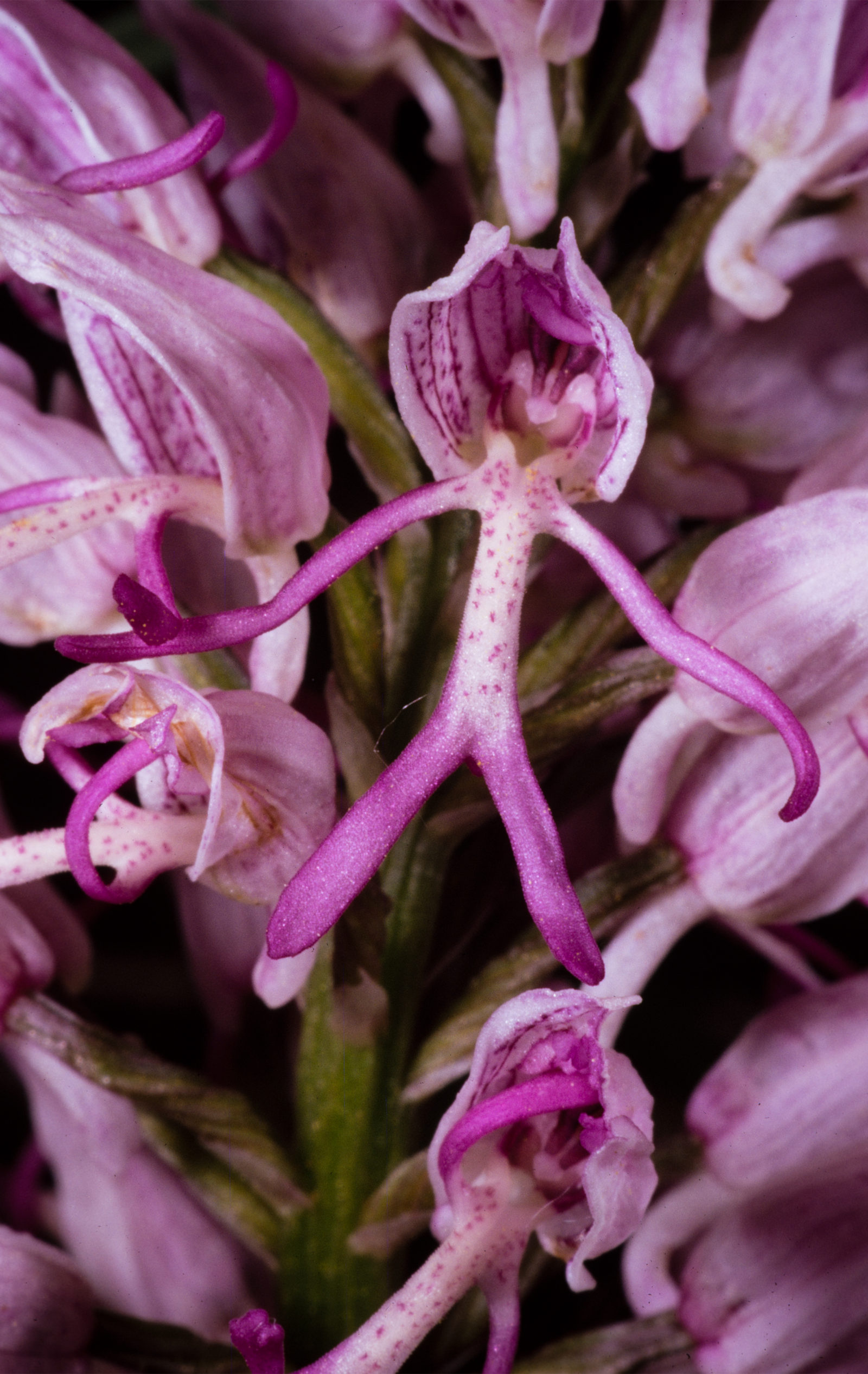
But these photos form only part of Reinhard’s orchid documentation. He also recorded his observations and findings in the form of some 1,800 plant and flower analyses. These include over 500 flower analyses, mounted in slide frames. The entire orchid documentation was placed in the ETH-Bibliothek archives via the ETH Zurich Geobotanical Institute. 11
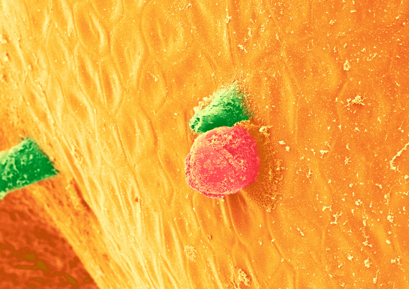
Beyond visibility – Plant microstructures in image form
Nowadays images created with electron microscopes play a vital role in the representation of plant microstructures. Yet the invention of the light microscope as early as the seventeenth century had for the first time enabled insights into the internal structure of plants.
Publications like Nehemia Grew’s ‘The Anatomy of Plants’ of 1682 contained detailed drawings created with the help of a microscope. In 1764, in his work ‘Das neueste aus dem Reiche der Pflanzen’, Johann Christoph Keller dedicated a richly illustrated chapter to the newly developed universal microscope. 12 But despite continuous improvements in optics, the smallest structures of plants remained invisible until well into the twentieth century.
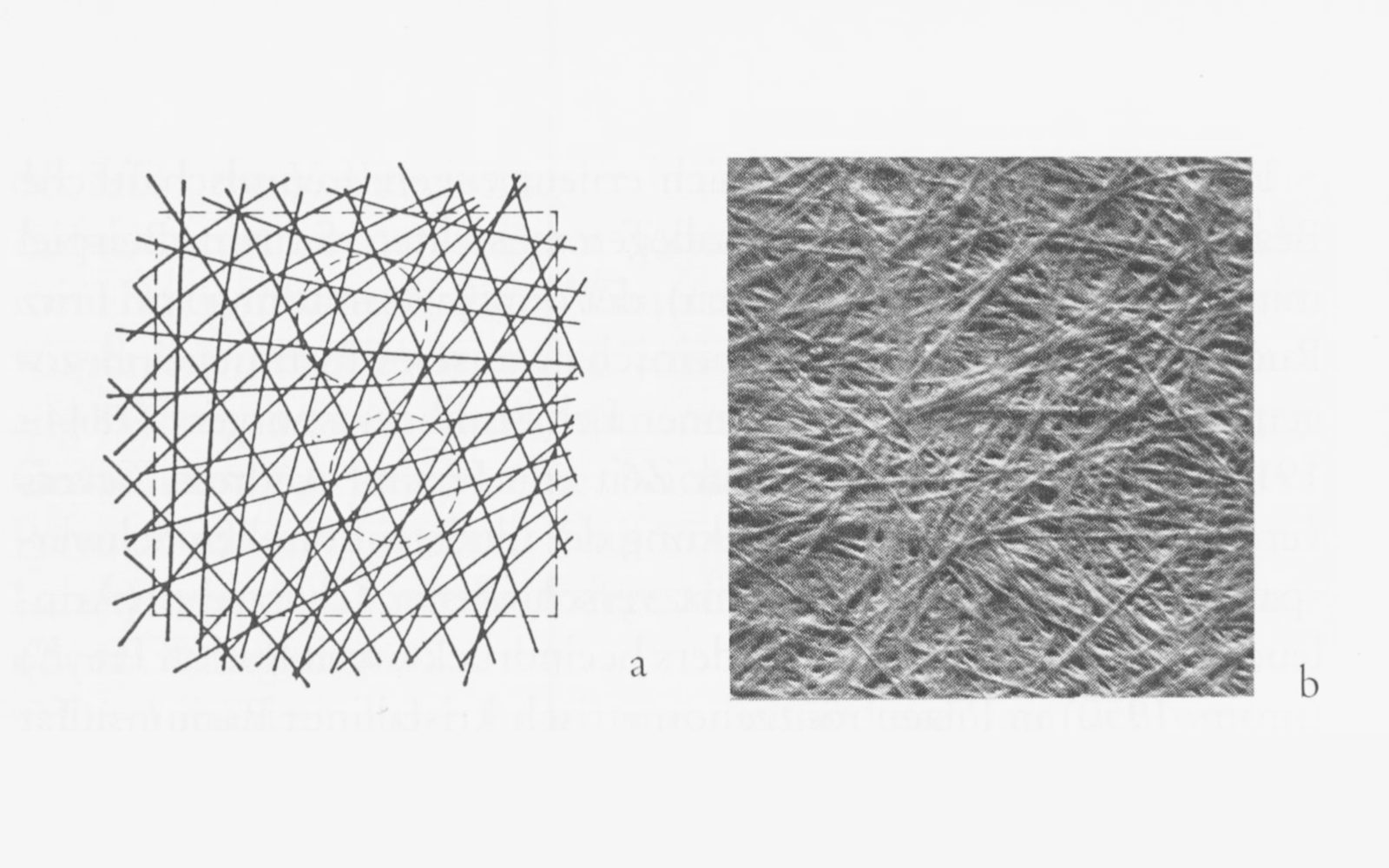
Thus in the 1930s at ETH Zurich, Albert Frey-Wyssling (1900–1988), professor of general botany and plant physiology, researched the submicroscopic structures of plants with indirect methods. For example, by analysing refractions he developed models – and drawings – of the internal structures of cell walls.
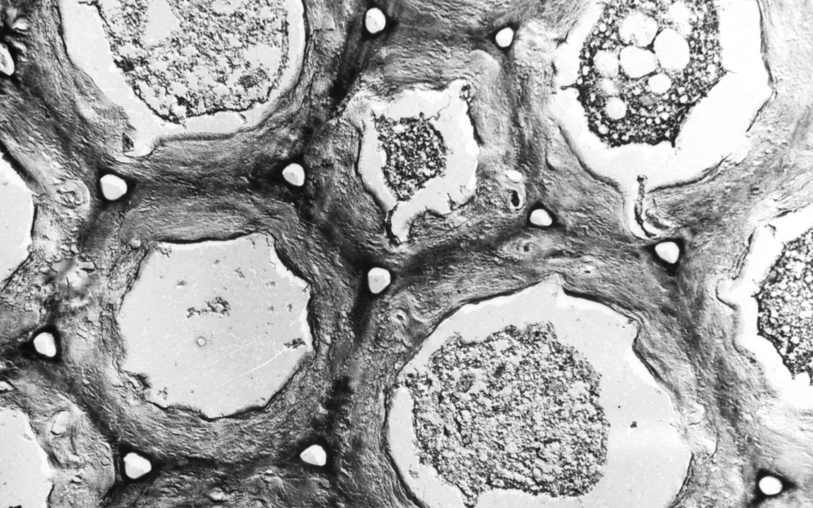
To promote his research in this area, Frey-Wyssling became heavily involved in the development of the electron microscope. From 1944 he established the first laboratory in Switzerland for electron microscopy at the ETH Zurich Institute for General Botany. It was with satisfaction that Frey-Wyssling found that the images generated by the new device provided visual confirmation of his earlier research:
It was clear that speculations of the [indirect] type mentioned could not lead to recognition of submicroscopic morphology. It was the electron microscope that led to the great breakthrough. It showed the correctness of the rod model of cell walls and the shift model of chloroplasts, on which these structures – opened up as a result of indirect methods – were suddenly presumed to be right because ‘it was now possible to see them’. 13
— Albert Frey-Wyssling, 1984
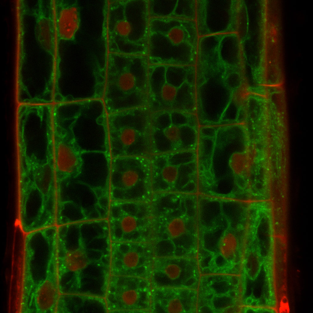
3D visualisations and confocal microscopy: The possibilities of modern technology
With digitisation, knowledge transfer is becoming – in visual terms – ever more attractive, more narrative and faster in the twenty-first century. Established representation techniques are being refined by the demands of specialised research. This also involves experimenting with new methods of representing plants. Specialist software, web applications and interactive representations are being used to depict scientific findings. Particularly popular in this respect are animated pictures, starting off, for example, with microscopic images or 3D models that make visible the dynamic processes inside and outside plants. Interactive 3D visualisations are the latest extension in the portfolio of scientific plant illustrations.

But modern methods also play a significant role beyond teaching and research in science communication, an area whose importance is growing all the time. Visual appeal has helped and is helping to reach a broad audience in addition to specialist groups and to make complex connections comprehensible to everyone. Use is being made in this respect of a growing palette of visualisation forms, often at an interface with artistic and creative endeavours.
The continuing exchange seen over the centuries between players from science, technology, art and design will also drive exciting developments in the range of visualisation opportunities available in the future. This is true not just in plant illustrations but also for scientific visualisations in general.
Footnotes
- Jacquin, Niklaus Joseph von: Florae austriacae sive plantarum selectarum in Austriae archiducatu sponte crescentium icones, Vienna: typis Leopoldi Joannis Kaliwoda, 1773–1778. ↩︎
- Reichenbach, Ludwig: Deutschlands Flora : mit höchst naturgetreuen, charakteristischen Abbildungen aller ihrer Pflanzen-Arten in natürlicher Grösse und mit Analysen auf Kupfertafeln als Belege für die Flora Germanica Excursoria, Leipzig: Friedrich Hofmeister, 1837–1867. ↩︎
- Labram, Jonas David; Hegetschweiler, Johannes: Sammlung von Schweizer Pflanzen nach der Natur und auf Stein gezeichnet, Zurich: Esslinger, o. D. ↩︎
- Schweizerischer Landwirtschaftlicher Verein (Hg.): Schweizerische Obstsorten, o. O.: Schweizerischer Landwirtschaftlicher Verein, 1863, Bd. 1, Preface. ↩︎
- Schweizerischer Landwirtschaftlicher Verein (Hg.), Schweizerische Obstsorten, o. O.: Schweizerischer Landwirtschaftlicher Verein, 1863, Bd. 2, Closing words. ↩︎
- Heer, Oswald: Flora Tertiaria Helvetiae = Die Tertiäre Flora Der Schweiz, Winterthur: Verlag der Lithographischen Anstalt von J. Wurster & Compagnie, 1855-1859, Bd. 1, vi. ↩︎
- https://www.google.ch/maps/place/focusTerra/@47.3785812,8.54503,17z/data=!3m1!4b1!4m5!3m4!1s0x479aa0a6ff9ea61d:0x9f67ca71c5f885dc!8m2!3d47.3785776!4d8.5472187 ↩︎
- https://www1.ethz.ch/collections.erdw/sammlung/geol/geol_historisch/heer_folder/Ohningen_im_Miozan.pdf ↩︎
- Hess, Hans Ernst; Landolt, Elias; Hirzel, Rosmarie: Flora der Schweiz und angrenzender Gebiete, second, revised edition, Basel: Birkhäuser, 1976, vol. 1, p. 15. ↩︎
- Baltisberger, Matthias; Nyffeler, Reto; Widmer, Alex: Systematische Botanik. Einheimische Farn- und Samenpflanzen, fourth edition, Zurich: v/d/f, 2013. ↩︎
- This extensive botanical collection has been accessible since May 2016: Plant analyses on the platform e-manuscripta and colour slides in the catalogue ETH E-Pics animals, plants and habitats. In 2016 the even bigger collection of images of European orchids by Peter Gölz, who often went on expeditions jointly with Hans Reinhard, was transferred to ETH Library, where it has also been made accessible. ↩︎
- Keller, Johann Christoph; Gleichen-Russwurm, Wilhelm Friedrich von: Das neueste aus dem Reiche der Pflanzen, Nürnberg: Gedruckt bei Christian de Launoy seel. Erben, 1764, pp. 9ff. ↩︎
- Frey-Wyssling, Albert. Lehre und Forschung: Autobiographische Erinnerungen. Volume 44. Grosse Naturforscher. Stuttgart: Wissenschaftliche Verlagsgesellschaft, 1984, pp. 87–88. ↩︎